SARS-CoV-2 coronavirus, antibodies, vaccines
Attacking the coronavirus

Transmission electron microscope image showing SARS-CoV-2
This transmission electron microscope image shows SARS-CoV-2—also known as 2019-nCoV, the virus that causes COVID-19—isolated from a patient in the U.S. Virus particles are shown emerging from the surface of cells cultured in the lab. The spikes on the outer edge of the virus particles give coronaviruses their name, crown-like.
Credit: NIAID-RML
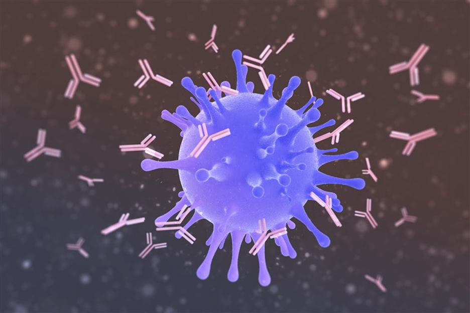
Computer illustration showing antibodies (pink) attacking a virus particle (blue)
Computer illustration showing antibodies (pink) attacking a virus particle (blue).
Transmission electron microscope image showing SARS-CoV-2
This transmission electron microscope image shows SARS-CoV-2—also known as 2019-nCoV, the virus that causes COVID-19—isolated from a patient in the U.S. Virus particles are shown emerging from the surface of cells cultured in the lab. The spikes on the outer edge of the virus particles give coronaviruses their name, crown-like.
Credit: NIAID-RML
Computer illustration showing antibodies (pink) attacking a virus particle (blue)
Computer illustration showing antibodies (pink) attacking a virus particle (blue).
Source: Getty Images
Antibodies
When pathogens like the coronavirus, SARS-CoV-2, attack, the body creates antibodies to neutralize the invader as part of the immune response.
An Antibody is a protein produced by the immune system in response to invading organisms such as bacteria and viruses. Antibodies are proteins produced by the immune system from the body’s stores of immunoglobulin protein. The immune system cells produce antibodies when they react with foreign protein antigens.
Antibodies are glycoproteins belonging to the immunoglobulin (Ig) superfamily. The terms antibody and immunoglobulin are often used interchangeably, though the term antibody is sometimes reserved for the secreted, soluble form which excludes B-cell receptors.
Antibodies are able to specifically recognize foreign invaders in the body, bind to them and then allow the immune cells of the body to clear them from the blood or mucous membranes, or sites like the lung. Each antibody can bind to only one specific foreign organism. Some destroy it directly; others make it easier for white blood cells to destroy it.
The immune response
The immune response includes initial and secondary antibodies:
• Immunoglobulin M, or IgM, is the initial antibody made to fight an invader
• Immunoglobulin A, or IgA, is typically found in saliva and other secretions
• Immunoglobulin G, or IgG, is the main type of antibody found in all body fluids
After initially making IgM, the cells switch over to making IgG, which is the longer-lived kind of antibody.
The presence of IgM means a person is currently infected or very recently became infected. Having IgG means the infection could have happened a month or two ago.
The adaptive immune system
Together with B and T cells, antibodies are the most important part of the adaptive immune system, also referred as the acquired immune system, which is a subsystem of the immune system that is composed of specialized, systemic cells and processes that eliminate pathogens or prevent their growth. The other main immunity strategy found in vertebrates is the innate immune system.
Soluble antibodies are released into the blood and tissue fluids, as well as many secretions. Because these fluids were traditionally known as humors, antibody-mediated immunity is sometimes known as or considered a part of humoral immunity.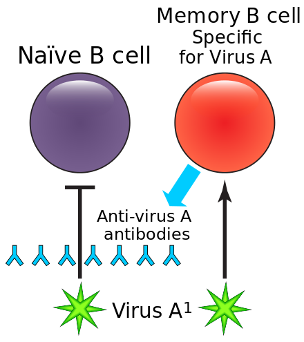
https://en.wikipedia.org/wiki/Memory_B_cell
Initially, antibodies are attached to the surface of a B cell and referred to as B-cell receptors (BCR). After an antigen binds to a BCR, the B cell activates to proliferate and differentiate into either plasma cells, which are white blood cells that originate in the bone marrow and secrete soluble antibodies with the same paratope, or memory B cells, which survive in the body to enable long-lasting immunity to the antigen.
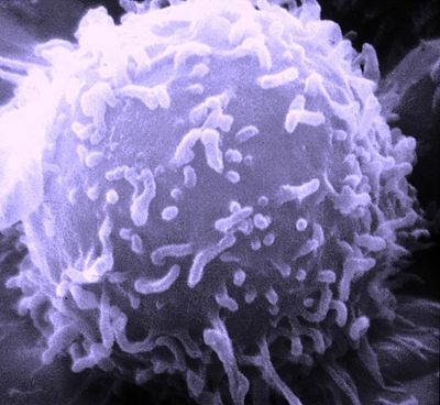
SEM image: single human lymphocyte.
https://en.wikipedia.org/wiki/Adaptive_immune_system
B lymphocytes are the cells of the immune system that make antibodies to invading pathogens like viruses. They form memory cells that remember the same pathogen for faster antibody production in future infections.
The antibody isotype of a B cell changes during cell development and activation. Immature B cells, which have never been exposed to an antigen, express only the IgM isotype in a cell surface bound form. The B lymphocyte, in this ready-to-respond form, is known as a “naive B lymphocyte.
The naive B lymphocyte expresses both surface IgM and IgD. The co-expression of both of these immunoglobulin isotypes renders the B cell ready to respond to antigen. B cell activation follows engagement of the cell-bound antibody molecule with an antigen, causing the cell to divide and differentiate into an antibody-producing cell called a plasma cell. In this activated form, the B cell starts to produce antibody in a secreted form rather than a membrane-bound form. Some daughter cells of the activated B cells undergo isotype switching, a mechanism that causes the production of antibodies to change from IgM or IgD to the other antibody isotypes, IgE, IgA, or IgG, that have defined roles in the immune system.
B cell receptors
The membrane-bound form of an antibody may be called a surface immunoglobulin (sIg) or a membrane immunoglobulin (mIg). It is part of the B cell receptor (BCR), which allows a B cell to detect when a specific antigen is present in the body and triggers B cell activation. The BCR is composed of surface-bound IgD or IgM antibodies and associated Ig-α and Ig-β heterodimers, which are capable of signal transduction.
A typical human B cell will have 50,000 to 100,000 antibodies bound to its surface. Upon antigen binding, they cluster in large patches, which can exceed 1 micrometer in diameter, on lipid rafts that isolate the BCRs from most other cell signaling receptors. These patches may improve the efficiency of the cellular immune response. In humans, the cell surface is bare around the B cell receptors for several hundred nanometers, which further isolates the BCRs from competing influences.
Proteins
Proteins are large biomolecules or macromolecules that are comprised of one or more long chains of amino acid residues. Proteins perform a vast array of functions within organisms, including catalysing metabolic reactions, DNA replication, responding to stimuli, providing structure to cells and organisms, and transporting molecules from one location to another. Proteins differ from one another primarily in their sequence of amino acids, which is dictated by the nucleotide sequence of their genes, and which usually results in protein folding into a specific 3D structure that determines its activity.
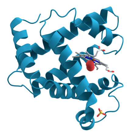
3D structure of myoglobin
A representation of the 3D structure of the protein myoglobin showing turquoise α-helices. This protein was the first to have its structure solved by X-ray crystallography. Toward the right-center among the coils, a prosthetic group called a heme group (gray) with a bound oxygen molecule (red). (https://en.wikipedia.org/wiki/Protein)
A linear chain of amino acid residues is called a polypeptide. A protein contains at least one long polypeptide. Short polypeptides, containing less than 20–30 residues, are rarely considered to be proteins and are commonly called peptides, or sometimes oligopeptides. The individual amino acid residues are bonded together by peptide bonds and adjacent amino acid residues. The sequence of amino acid residues in a protein is defined by the sequence of a gene, which is encoded in the genetic code. In general, the genetic code specifies 20 standard amino acids; but in certain organisms the genetic code can include selenocysteine and—in certain archaea—pyrrolysine.
Antibody structure; in general
An antibody (Ab) is a large, Y-shaped protein used by the immune system to identify and neutralize foreign objects such as pathogenic bacteria and viruses.
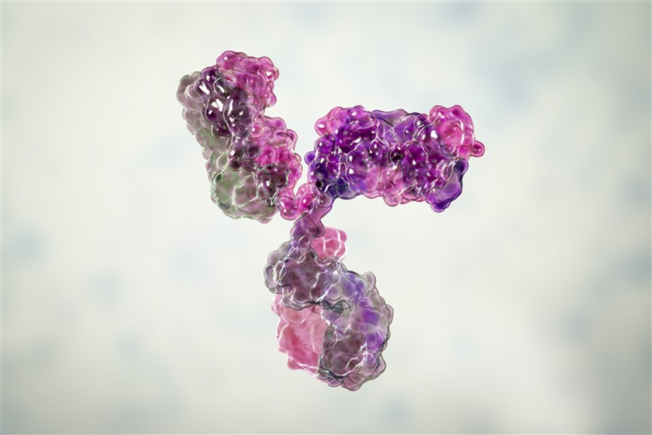
immunoglobulin G antibody molecule: a computer model. Getty Images
A computer model shows the immunoglobulin G antibody molecule. Each Y-shaped molecule has two arms (top) that can bind to bacterial or viral proteins marking the virus for destruction by white blood cells.
The antibody recognizes a unique molecule of the pathogen, called an antigen (Ag) which is a molecule or molecular structure, such as may be present on the outside of a pathogen, that can be bound by an antigen-specific antibody or B-cell antigen receptor. The presence of antigens in the body normally triggers an immune response. The Ag abbreviation stands for an antibody generator.
Antigens are “targeted” by antibodies. Each antibody is specifically produced by the immune system to match an antigen after cells in the immune system come into contact with it which allows a precise identification or matching of the antigen and the initiation of an adaptive response.
Antigens are proteins, peptides (amino acid chains) and polysaccharides (chains of monosaccharides/simple sugars) but lipids and nucleic acids become antigens only when combined with proteins and polysaccharides.
Each tip of the “Y” of an antibody contains a paratope or antigen binding site analogous to a lock that is specific for one particular epitope or antigenic determinant analogous to a key on an antigen that is recognized by the immune system, specifically by antibodies, B cells, or T cells.
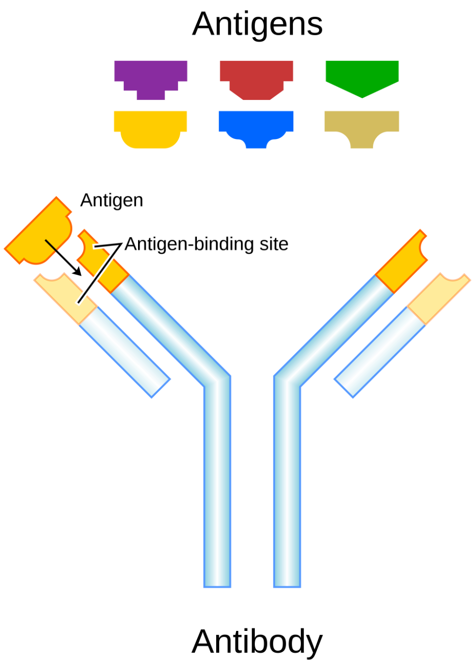
Antigen (key) and antibody (lock) model
Vaccines are examples of antigens in an immunogenic form, which are intentionally administered to a recipient to induce the memory function of the adaptive immune system towards antigens of the pathogen invading that recipient.
Antibody structure; details
In humans and most mammals, an antibody unit consists of four polypeptide chains; two identical heavy chains and two identical light chains connected by disulfide bonds. Each chain is a series of domains: somewhat similar sequences of about 110 amino acids each. These domains are usually represented in simplified schematics as rectangles. Light chains consist of one variable domain VL and one constant domain CL, while heavy chains contain one variable domain VH and three to four constant domains CH1, CH2, …
The basic unit of an antibody, or immunoglobulin, molecule. is composed of two identical light (L) chains and two identical heavy (H) chains, which are held together by disulfide bonds to form a flexible Y shape. Each chain is composed of a variable (V) region and a constant (C) region. The soluble Y-shaped units can occur individually as monomers, or in complexes of two to five units.

antibody structural schematic
Schematic structure of an antibody: two heavy chains (blue, yellow) and the two light chains (green, pink). The antigen binding site is circled.
Structurally an antibody is also partitioned into two antigen-binding fragments (Fab), containing one VL, VH, CL, and CH1 domain each, as well as the crystallisable fragment (Fc), forming the trunk of the Y shape. In between them is a hinge region of the heavy chains, whose flexibility allows antibodies to bind to pairs of epitopes at various distances, to form complexes (dimers, trimers, etc.), and to bind effector molecules more easily.
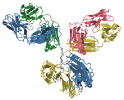
antibody 3-D structure at RCSB PDB.
Antibodies are heavy (~150 kDa) proteins of about 10 nm in size, arranged in three globular regions that roughly form a Y shape.
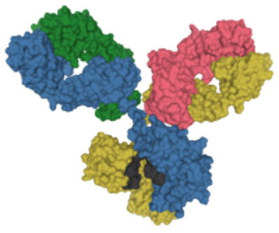
antibody 3-D structure at RCSB PDB.
Glycans in the Fc region are shown in black.
The antigen-binding sites at both tips of the antibody come in a wide variety while the remainder of the antibody is relatively constant with only a few variants which define the antibody’s class or isotype: IgA, IgD, IgE, IgG, or IgM. The constant region at the trunk of the antibody includes sites involved in interactions with other components of the immune system. The class hence determines the function triggered by an antibody after binding to an antigen in addition to some structural features.
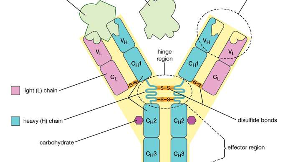
The 4 chain structure of an antibody. (Encyclopædia Britannica, Inc.)
Antibody classes or idiotypes
The five antibody classes of placental mammals are further subdivided into subclasses such as IgA1, IgA2. The prefix “Ig” stands for immunoglobulin, while the suffix denotes the type of heavy chain the antibody contains: the heavy chain types α (alpha), γ (gamma), δ (delta), ε (epsilon), μ (mu) give rise to IgA, IgG, IgD, IgE, IgM, respectively. The distinctive features of each class are determined by the part of the heavy chain within the hinge and Fc region.
The classes differ in their biological properties, functional locations and ability to deal with different antigens. For example, IgE antibodies are responsible for an allergic response consisting of histamine release from mast cells, contributing to asthma. The antibody’s variable region binds to allergic antigen, for example house dust mite particles, while its Fc region (in the ε heavy chains) binds to Fc receptor ε on a mast cell, triggering its degranulation: the release of molecules stored in its granules.
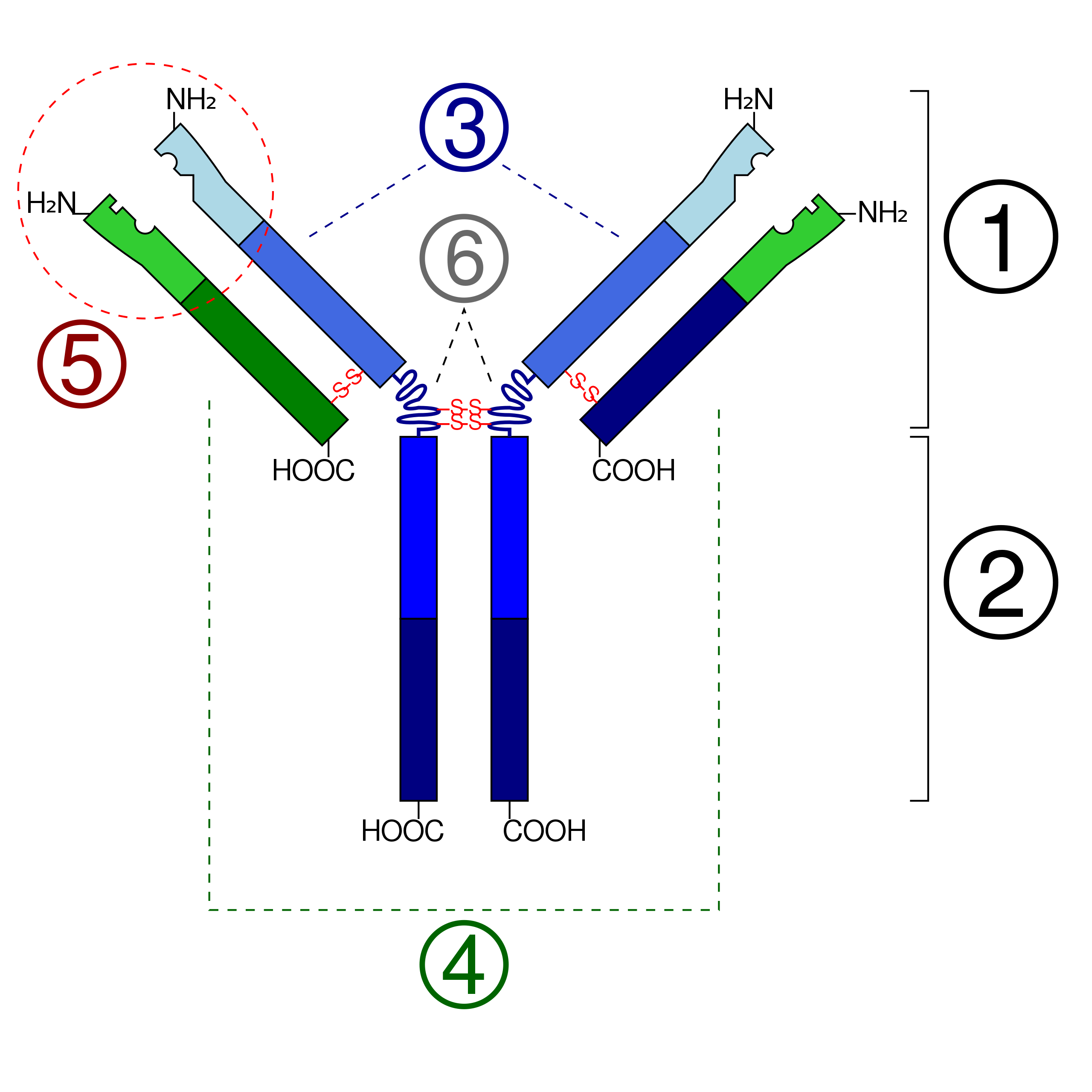
Antibody schematic; circled region depicts paratope location
1. Antigen-binding fragment (Fab) 2. Antibody crystallizable region (Fc) 3. Heavy chains 4. Light chains 5. Variable region of the antibody. 6. Hinge regions.
The paratope is the key-shaped section that makes direct contact with the antigen.
Antibody complexes
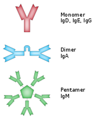
Antibody complexes
Secreted antibodies can occur as a single Y-shaped unit, a monomer. However, some antibody classes also form dimers with two Ig units (as with IgA), tetramers with four Ig units (like teleost fish IgM), or pentamers with five Ig units (like mammalian IgM, which occasionally forms hexamers as well, with six units).
Antibodies also form complexes by binding to antigen: this is called an antigen-antibody complex or immune complex. Small antigens can cross-link two antibodies, also leading to the formation of antibody dimers, trimers, tetramers, etc. Multivalent antigens (cells with multiple epitopes) can form larger complexes with antibodies. An extreme example is the clumping, or agglutination, of red blood cells with antibodies in the Coombs test to determine blood groups: the large clumps become insoluble, leading to visually apparent precipitation.
Vaccines; in general
Viruses are intracellular parasites that need host cells to multiply. Thus, for a virus to infect a human, it has to get access to some of the body’s cells that will enable viruses to multiply. Progeny viruses will be assembled within the infected cells and, upon release, will infect other surrounding target cells. Without any immune response to counteract the virus, it will continue to spread and cause organ damage, disease, and eventual death.
Vaccines are the most cost-effective way to provide a host with virus-specific immunity that will then help it keep an infectious virus below pathogenic levels. To accomplish this, vaccines may induce antibodies that help to neutralize assembled free viruses and virus-specific cytotoxic T cells that will kill infected cells and thus reduce the number of virus-producing cells.
While both elements of the immune response are considered of major importance for vaccine efficacy, the question is how do they cooperate? Researchers have now addressed these fundamental questions by examining the contribution of antibodies and cytotoxic T cells using a mathematical model based on virus infection dynamics. They show that these two primary control factors of virus infection are cooperating multiplicatively rather than additively.